Home
/ Bones In Your Leg Diagram : The Leg Ankle And Foot Amboss : Watch this 'leg bones' video lesson to discover all about the leg bones.
Bones In Your Leg Diagram : The Leg Ankle And Foot Amboss : Watch this 'leg bones' video lesson to discover all about the leg bones.
Bones In Your Leg Diagram : The Leg Ankle And Foot Amboss : Watch this 'leg bones' video lesson to discover all about the leg bones.. The femur, or thigh bone, is the largest, heaviest, and strongest bone in the human body. This diagram of a human skeleton is labeled with 12 major bones, from skull to fibula. At the distal end of the femur, two rounded condyles meet the tibia and fibula bones of the lower leg to form the knee joint. Posted on january 20, 2015 by admin. A diagram of the human skeleton showing bone and cartilage.
When your muscles contract, they pull the bone they're. Located in the upper leg, the thigh bone is known as the femur; License image the bones of the leg are the femur, tibia, fibula and patella. Learn vocabulary, terms and more with flashcards, games and other study tools. But if something goes a bit wrong, they can hurt and make it hard to move around.
Tibia And Fibula Anatomy Of Leg Bones Anatomy Physiology Youtube from i.ytimg.com Normal leg bones are relatively straight, but those affected by paget's disease are porous and figure 9. The longest bone in the human is called the. You can specify conditions of storing and accessing cookies in your browser. It allows us to walk, run, and jump. When your muscles contract, they pull the bone they're. A diagram of the human skeleton showing bone and cartilage. This diagram of a human skeleton is labeled with 12 major bones, from skull to fibula. , knee, and leg pain helps you understand how trigger points form and where to search for them.
Want to learn more about bones?
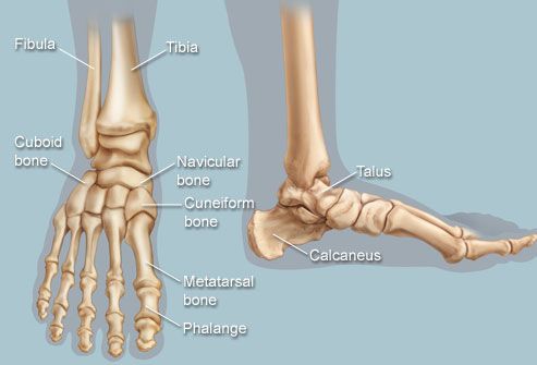
License image the bones of the leg are the femur, tibia, fibula and patella. This diagram of a human skeleton is labeled with 12 major bones, from skull to fibula. The human body is to some extent like a walking tower that moves on pillars, represented by the legs. The bones of the leg are the femur, tibia, fibula and patella. The talus bone supports the leg bones (tibia and fibula), forming the ankle. I had to reverse some numbers because the model i was working with has bird legs, but as soon as i figured that out this was immensely helpful. Pencil drawing on paper one of a kind artwork size: The foot bones shown in this diagram are the talus, navicular, cuneiform, cuboid, metatarsals and calcaneus. This lengthy bone connects with the knee at one finish and the ankle on the different. D) that the shape of the bones has less to do with the environment. Of one blender unit of length. Learn vocabulary, terms and more with flashcards, games and other study tools. Your leg bones are the longest and strongest bones in your body.
Your leg bones are very large and strong to help support the weight of your body. Located in the upper leg, the thigh bone is known as the femur; The foot bones shown in this diagram are the talus, navicular, cuneiform, cuboid, metatarsals and calcaneus. Continue scrolling to read more below. The knee is a strong but flexible hinge joint.
Feet Human Anatomy Bones Tendons Ligaments And More from img.webmd.com Download this free vector about diagram showing the hip bone treatment, and discover more than 15 million professional graphic resources on freepik. Continue scrolling to read more below. C) that they developed their bone structure independently of one another. A diagram of the human skeleton showing bone and cartilage. I had to reverse some numbers because the model i was working with has bird legs, but as soon as i figured that out this was immensely helpful. Normal leg bones are relatively straight, but those affected by paget's disease are porous and figure 9. 11.02 x 15.75 in (unframed). Your leg bones are very large and strong to help support the weight of your body.
The femur, or thigh bone, is the largest, heaviest, and strongest bone in the human body.
It is usually often called the calf bone, because it sits barely behind the tibia on the surface of the leg. The head of the femur rests inside the acetabulum in the pelvic bone; Located in the upper leg, the thigh bone is known as the femur; I had to reverse some numbers because the model i was working with has bird legs, but as soon as i figured that out this was immensely helpful. However, the definition in human anatomy refers only to the foot bones shown in this diagram are the talus, navicular, cuneiform, cuboid, metatarsals and calcaneus. The knee is a strong but flexible hinge joint. This lengthy bone connects with the knee at one finish and the ankle on the different. Normal leg bones are relatively straight, but those affected by paget's disease are porous and figure 9. You can specify conditions of storing and accessing cookies in your browser. The lower limb in the modern human is an interestingly adapted limb to bipedal walking, and as such it has changed anatomically from our nearest cousins growth of the long bones in a juvenile knee joint (the femur is located proximally, with tibia distal and fibula laterally. What does this suggest about mammals? Posted on january 20, 2015 by admin. When you stand or walk, all the weight of your upper body rests on them.
The most common cause is diabetes, but other health conditions, medicines, injuries, or infections can cause it. That nourish every function of the body, and it is the bone marrow that nourishes and rejuvenates the org. When you stand or walk, all the weight of your upper body rests on them. With its root placed at the 3d cursor position. Tremendous advantages have been gained from this erect posture, the chief among which has been.
8 4 Bones Of The Lower Limb Anatomy Physiology from open.oregonstate.education All those exercises that stretch you feet and leg muscles are good for flat feet. Located in the upper leg, the thigh bone is known as the femur; The knee is a strong but flexible hinge joint. The most common cause is diabetes, but other health conditions, medicines, injuries, or infections can cause it. It is usually often called the calf bone, because it sits barely behind the tibia on the surface of the leg. But if something goes a bit wrong, they can hurt and make it hard to move around. Of one blender unit of length. , knee, and leg pain helps you understand how trigger points form and where to search for them.
Your legs are an amazing collection of bones and muscles.
Editor · aug 13, 2017 ·. The lower limb has 30 bones some of which are tibia, femur, tarsal bones, fibula, metatarsal bones, etc. The talus bone supports the leg bones (tibia and fibula), forming the ankle. It is the bone closest to the center of the body. The foot bones shown in this diagram are the talus, navicular, cuneiform, cuboid, metatarsals and calcaneus. Just like the three bones in the arm, the three. Tremendous advantages have been gained from this erect posture, the chief among which has been. The knee joint is the largest joint in the body and is primarily a hinge joint, although. What does this suggest about mammals? The femur, or thigh bone, is the largest, heaviest, and strongest bone in the human body. Stress and leg weakness are detrimental to broiler production, health, and welfare. At the distal end of the femur, two rounded condyles meet the tibia and fibula bones of the lower leg to form the knee joint. That nourish every function of the body, and it is the bone marrow that nourishes and rejuvenates the org.

